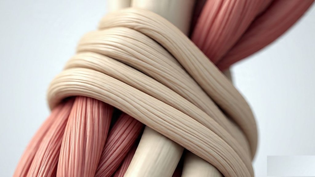
In the intricate architecture of the human body, muscles do not work in isolation. They rely on specialized connective tissues to anchor them to bones or other tissues. One of the most essential and often overlooked structures in this system is the aponeurosis—a flat sheet or ribbon-like structure made of tendon-like material. This crucial tissue serves as a bridge between muscles and the parts they move, enabling coordinated and efficient motion.
In this article, we’ll explore what an aponeurosis is, its structure, functions, types, and clinical significance.
What Is an Aponeurosis?
An aponeurosis (plural: aponeuroses) is a broad, flat sheet of dense fibrous connective tissue. Like tendons, aponeuroses anchor muscles to bones or other tissues. However, while tendons are cord-like, aponeuroses are sheet-like, providing a wider surface area for muscle attachment.
They play a vital role in transmitting the force generated by a muscle to the skeletal system or other muscular structures. This function makes them indispensable in many regions of the body where broad muscle attachment is required.
Structure of an Aponeurosis
Aponeuroses are composed of dense regular connective tissue, which provides both strength and flexibility. Key components include:
1. Fibroblasts
These are spindle-shaped, collagen-producing cells. In aponeuroses, fibroblasts are relatively sparse but essential—they synthesize and maintain the extracellular matrix, particularly collagen.
2. Collagenous Fibers
The bulk of an aponeurosis is formed by bundles of collagen fibers arranged in orderly, parallel arrays. This arrangement provides high tensile strength, allowing the aponeurosis to withstand significant pulling forces.
3. Elastic Fibers (in smaller quantities)
Some aponeuroses also contain elastic fibers, which provide a degree of elasticity, although their role is much less prominent than that of collagen.
Function of the Aponeurosis
The primary function of the aponeurosis is to connect muscles to the parts they move. Additional roles include:
- Force Transmission: Aponeuroses act as force transmitters, helping convey muscle contraction to bones or other structures.
- Muscle Support: In areas where muscles lack direct bony attachment, aponeuroses provide broad anchoring support.
- Structural Reinforcement: In certain regions, aponeuroses help reinforce the integrity of body walls or cavities.
- Pressure Distribution: By spreading muscle forces across a larger area, they help avoid localized pressure or injury.
Examples of Aponeuroses in the Human Body
1. Palmar Aponeurosis
Located in the palm of the hand, this structure connects the tendons of the forearm muscles to the skin and fascia of the palm. It assists with gripping and protects underlying nerves and vessels.
2. Plantar Aponeurosis
Also known as the plantar fascia, this thick aponeurosis runs along the sole of the foot. It supports the arch and aids in the biomechanics of walking.
3. Abdominal Aponeuroses
The flat muscles of the abdominal wall (external oblique, internal oblique, and transversus abdominis) end in broad aponeuroses that meet at the linea alba, a central fibrous seam.
4. Epicranial Aponeurosis (Galea Aponeurotica)
This is the fibrous sheet connecting the frontalis muscle (on the forehead) to the occipitalis muscle (on the back of the head). It plays a role in facial expression and scalp movement.
Differences Between Tendons and Aponeuroses
| Feature | Tendon | Aponeurosis |
| Shape | Cord-like | Sheet-like |
| Function | Muscle-to-bone attachment | Muscle-to-bone or muscle-to-muscle attachment |
| Structure | Dense collagen bundles | Dense collagen, broad arrays |
| Example | Achilles tendon | Palmar aponeurosis |
Clinical Significance
Understanding the structure and function of aponeuroses is essential for clinicians, surgeons, and physical therapists. Here are some medical conditions and considerations:
1. Plantar Fasciitis
Inflammation of the plantar aponeurosis (fascia) causes heel pain, commonly due to overuse, poor footwear, or abnormal gait mechanics.
2. Dupuytren’s Contracture
This involves the thickening and tightening of the palmar aponeurosis, causing fingers to bend inward and reducing hand function.
3. Abdominal Wall Hernias
Weakness or tearing in the abdominal aponeuroses can lead to hernias, where abdominal contents push through weakened tissues.
4. Surgical Reconstruction
In reconstructive surgeries, aponeuroses may be used as graft materials or may need to be carefully preserved to maintain function.
Evolutionary and Functional Adaptations
Aponeuroses have evolved in both humans and animals to meet specific biomechanical demands:
- In animals such as horses, specialized aponeuroses store and release energy, improving locomotive efficiency.
- In humans, their broad, flat nature allows for distributed force application, essential for maintaining posture and movement coordination.
Aponeurosis in Research and Biomechanics
Recent advances in biomechanical modeling and tissue engineering have renewed interest in aponeuroses:
- MRI and ultrasound help visualize aponeuroses in living tissues.
- Synthetic aponeurosis research aims to develop biomaterials for tissue repair.
- Biomechanical simulations now include aponeuroses to better predict human motion and design better prosthetics and exoskeletons.
Conclusion
Aponeuroses may not get the spotlight that bones and muscles do, but they are crucial components of the musculoskeletal system. Acting as silent facilitators, these flat sheets of connective tissue provide the strength, flexibility, and stability needed for a wide range of human movements.
From the arch of your foot to the frown on your brow, aponeuroses are at work. Understanding their form and function not only enriches our knowledge of human anatomy but also informs medical practice, athletic training, and even surgical innovation.
FAQs
Q1: Is aponeurosis the same as fascia?
No. While both are connective tissues, aponeuroses connect muscles to bones or other muscles, whereas fascia primarily wraps around muscles and organs to provide support.
Q2: Can aponeuroses regenerate after injury?
Mild injuries may heal with time and therapy. Severe damage may require surgical intervention, although healing can be slow due to limited blood supply.
Q3: Are aponeuroses found in all muscles?
No, aponeuroses are primarily found in flat muscles where broad surface attachment is needed.
Q4: What is the linea alba?
It is a central fibrous line in the abdomen formed by the joining of abdominal aponeuroses. It’s a landmark for surgeons and is often used in abdominal incisions.
Q5: Can I strengthen my aponeuroses through exercise?
While aponeuroses themselves don’t strengthen like muscles, regular training can improve the coordination and tension within the connected muscle-aponeurosis unit.

Theo Louse
I am Theo Louse. My skills are dedicated to the field of technology information and try to make daily lives more enjoyable. With more than 12 years of experience with BM, we are particularly famous for 100% self-developed ideas. Over these years, we have worked to make everyday life more convenient for the fast-paced world we live in.

